
Understanding the parts and functions of a microscope is essential for proper usage in education, research, and medicine. This guide explores key components, including the eyepiece, objective lenses, and stage, to enhance your microscopy skills and knowledge.
Overview of the Microscope
A microscope is an essential scientific tool designed to magnify small objects or specimens for detailed observation. It consists of a combination of optical, structural, and mechanical parts that work together to provide a clear and enlarged view of microscopic structures. The microscope is widely used in biology, medicine, and research to study cells, tissues, and microorganisms. Its basic operation involves focusing light through lenses to produce an image of the specimen. Understanding the microscope’s components and their functions is crucial for effective use in various scientific applications. This guide provides a comprehensive overview of the microscope’s parts, their roles, and how they contribute to its functionality, helping users gain proficiency in microscopy techniques.
Importance of Understanding Microscope Components
Understanding the components of a microscope is vital for maximizing its utility and ensuring accurate observations. Each part plays a specific role in achieving optimal magnification and clarity. Without knowledge of the eyepiece, objective lenses, stage, and other elements, users may struggle with proper specimen preparation and focusing. Familiarity with the microscope’s structure enhances experimental efficiency and reduces the risk of damage to the instrument. In educational and professional settings, comprehension of these components is essential for conducting precise analysis and interpreting results effectively. This knowledge also fosters better maintenance and troubleshooting, prolonging the microscope’s lifespan. Investing time in learning about these parts ensures a more rewarding and productive experience in microscopy.
Optical Parts of the Microscope
The optical parts, including the eyepiece, objective lenses, and condenser, work together to magnify and focus light, enabling clear visualization of specimens under examination.
Eyepiece (Ocular Lens)
The eyepiece, or ocular lens, is the lens through which the user views the specimen. Typically located at the top of the microscope, it magnifies the image by 10x. In binocular microscopes, two eyepieces are provided, one for each eye, ensuring a three-dimensional view. The eyepiece works in conjunction with the objective lenses to produce a magnified image. Proper alignment and adjustment of the eyepiece are crucial for clear observation. It is essential to focus the eyepiece correctly, especially for users with vision impairments, by using the diopter adjustment. This component is vital for optimizing the viewing experience and achieving accurate results in microscopy.
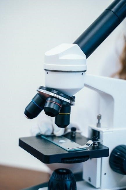
Objective Lenses
Objective lenses are crucial components mounted on the rotating nosepiece of a microscope. They are designed to focus light from the specimen and produce a magnified image. Common magnifications include 4x, 10x, 40x, and 100x, with higher magnifications used for detailed observations. Each lens is selected based on the desired level of detail and resolution. The objective lens collects light from the specimen and relays it to the eyepiece, which further magnifies the image. Proper alignment and focus of the objective lenses are essential for clear and accurate viewing. They are interchangeable, allowing users to switch between different magnifications easily. Objective lenses are fundamental to the functionality of a microscope, enabling users to study specimens at various scales with precision and clarity.
Condenser
The condenser is a critical component located below the stage of the microscope. Its primary function is to focus and direct light from the illuminator onto the specimen, ensuring even illumination. The condenser consists of a lens system that can be adjusted vertically to optimize light transmission. Proper alignment of the condenser is essential for achieving a sharp, clear image. It works in conjunction with the iris diaphragm to regulate the amount of light that reaches the specimen. Adjusting the condenser helps to improve contrast and resolution, making it easier to observe fine details of the specimen. Regular maintenance and alignment of the condenser are necessary to maintain the microscope’s performance and ensure accurate observations.
Structural Parts of the Microscope
The microscope’s structural framework includes the arm, base, and body tube. These components provide stability, support, and alignment for the optical and mechanical parts, ensuring precise functionality.
Arm
The arm is a sturdy metal component that connects the microscope’s body tube to the base. It provides structural support and serves as a bridge between the optical and mechanical parts. The arm allows the microscope to maintain balance and stability during use. Its durable design ensures that the microscope remains upright and secure, preventing any wobbling or movement that could disrupt observation. Additionally, the arm facilitates easy transportation of the microscope, as it offers a convenient handle for lifting and carrying the device. Proper alignment and strength of the arm are crucial for maintaining the microscope’s functionality and ensuring accurate observations. Without a robust arm, the microscope might not perform optimally, making it an indispensable structural component.
Base
The base is the foundational part of the microscope, providing stability and support for the entire instrument. It is typically made of durable materials such as metal or heavy-duty plastic to ensure balance and prevent movement during use. The base houses the illuminator, which is the light source used to illuminate specimens, and often includes a diaphragm to control light intensity. Its sturdy design ensures that the microscope remains stationary, which is critical for clear and precise observations. The base also serves as a platform for mounting other essential components, such as the arm and body tube; Proper construction of the base is vital to maintain the microscope’s functionality and performance, making it a crucial structural element in microscopy.
Body Tube
The body tube, also known as the microscope tube, is a cylindrical structure that connects the eyepiece to the objective lenses. It plays a crucial role in maintaining the optical alignment necessary for clear image formation. The body tube houses the internal optical system and ensures that light from the specimen travels correctly through the lenses. Its length and design are standardized to maintain proper magnification and focus. In some microscopes, the body tube may be adjustable to accommodate different eyepieces or objective lenses. Proper care of the body tube is essential to prevent dust and debris from interfering with the optical path, ensuring optimal performance during observations. The body tube’s robust construction also contributes to the overall durability of the microscope, making it a vital component in microscopy.

Mechanical Parts of the Microscope
The mechanical parts include the stage, stage clips, and focus knobs, which position and secure the specimen for viewing. These components ensure precise sample alignment and adjustment.
Stage
The stage is a flat platform located at the base of the microscope where the specimen slide is placed. It is typically made of metal or plastic and is designed to hold the slide securely in position. Some microscopes feature a mechanical stage, which has adjustable knobs that allow for precise movement of the slide in both the x and y directions. This feature is particularly useful for examining different areas of the specimen without having to remove the slide from the microscope. The stage often includes a slide holder or stage clips to keep the slide in place, ensuring stability and focus during observation. Proper alignment and security of the slide on the stage are crucial for obtaining clear and accurate images under magnification.
Stage Clips
Stage clips are small metal or plastic clamps located on the stage of the microscope. Their primary function is to securely hold the specimen slide in place during observation. These clips ensure that the slide remains stable, preventing any movement that could disrupt the focus or clarity of the image. Typically, there are two clips that grip the edges of the slide, and they can be adjusted to accommodate different slide sizes. Properly securing the slide with stage clips is essential for smooth operation, as it allows the user to focus and adjust the stage position without worrying about the slide shifting. This simple yet crucial component ensures that the specimen remains in the correct position for accurate and effective microscopy. Stage clips are an integral part of maintaining the integrity of the specimen preparation process.
Coarse and Fine Adjustment

The coarse and fine adjustments are critical focusing mechanisms in a microscope. The coarse adjustment knob moves the stage up or down in larger increments, bringing the specimen into approximate focus. Once the image is nearly clear, the fine adjustment knob is used to make smaller, precise movements, refining the focus for optimal clarity. These controls are essential for achieving sharp, detailed images of the specimen. Proper use of both adjustments ensures that the microscope maintains focus efficiently, preventing damage to the instrument or the slide. The coarse adjustment is typically used for initial focusing, while the fine adjustment fine-tunes the image. Together, they enable precise control over the microscope’s focus, making them indispensable for accurate observations. Proper handling of these adjustments is vital for effective microscopy.
Additional Components of the Microscope
The diaphragm regulates light intensity, while the illuminator provides the light source. Both components work together to optimize specimen visibility and image clarity during observation.
Diaphragm
The diaphragm is a crucial component located below the stage of the microscope. Its primary function is to regulate the amount of light passing through the specimen. By adjusting the diaphragm, users can control the intensity of light, ensuring optimal illumination for clear observation. It typically consists of a rotating disk with different-sized apertures, allowing for precise light modulation. The diaphragm works in conjunction with the condenser to focus light effectively. Proper use of the diaphragm enhances image clarity and contrast, making it essential for achieving accurate microscopic observations. It is particularly useful when working with specimens that require varying light levels for detailed analysis. Understanding how to adjust the diaphragm is a fundamental skill in microscopy, as it directly impacts the quality of the visual output.
Illuminator
The illuminator is a critical component of a microscope, serving as the light source that illuminates the specimen. Typically located at the base of the microscope, it consists of a light bulb, often LED or halogen, and a power supply. The illuminator’s primary function is to provide bright, even light to the condenser, which then focuses it onto the specimen. Many microscopes include an adjustable intensity control, such as a rheostat, allowing users to regulate light levels. Proper adjustment of the illuminator is essential for achieving clear and detailed images. It works in conjunction with the condenser and diaphragm to optimize light transmission, ensuring optimal visibility of the specimen under observation. Understanding how to use the illuminator effectively is fundamental for obtaining high-quality microscopic results.
A microscope’s effectiveness relies on understanding its components. Key parts include the eyepiece, objective lenses, stage, and condenser, each playing a vital role in enhancing observation and clarity.
Understanding the microscope’s components is crucial for effective use. The eyepiece magnifies images, while objective lenses provide varying magnification levels. The stage holds specimens securely, aided by stage clips. The arm supports the microscope’s body, connecting it to the base, which provides stability. The condenser focuses light onto the specimen, and the diaphragm adjusts light intensity. Coarse and fine adjustments enable precise focus. These parts work together to enhance observation and clarity, making microscopy essential in scientific and educational settings. Proper knowledge of these components ensures accurate and efficient use of the microscope for exploring microscopic structures.